
Dog ACL Tear: Signs, Surgery Options & What to Expect During Recovery
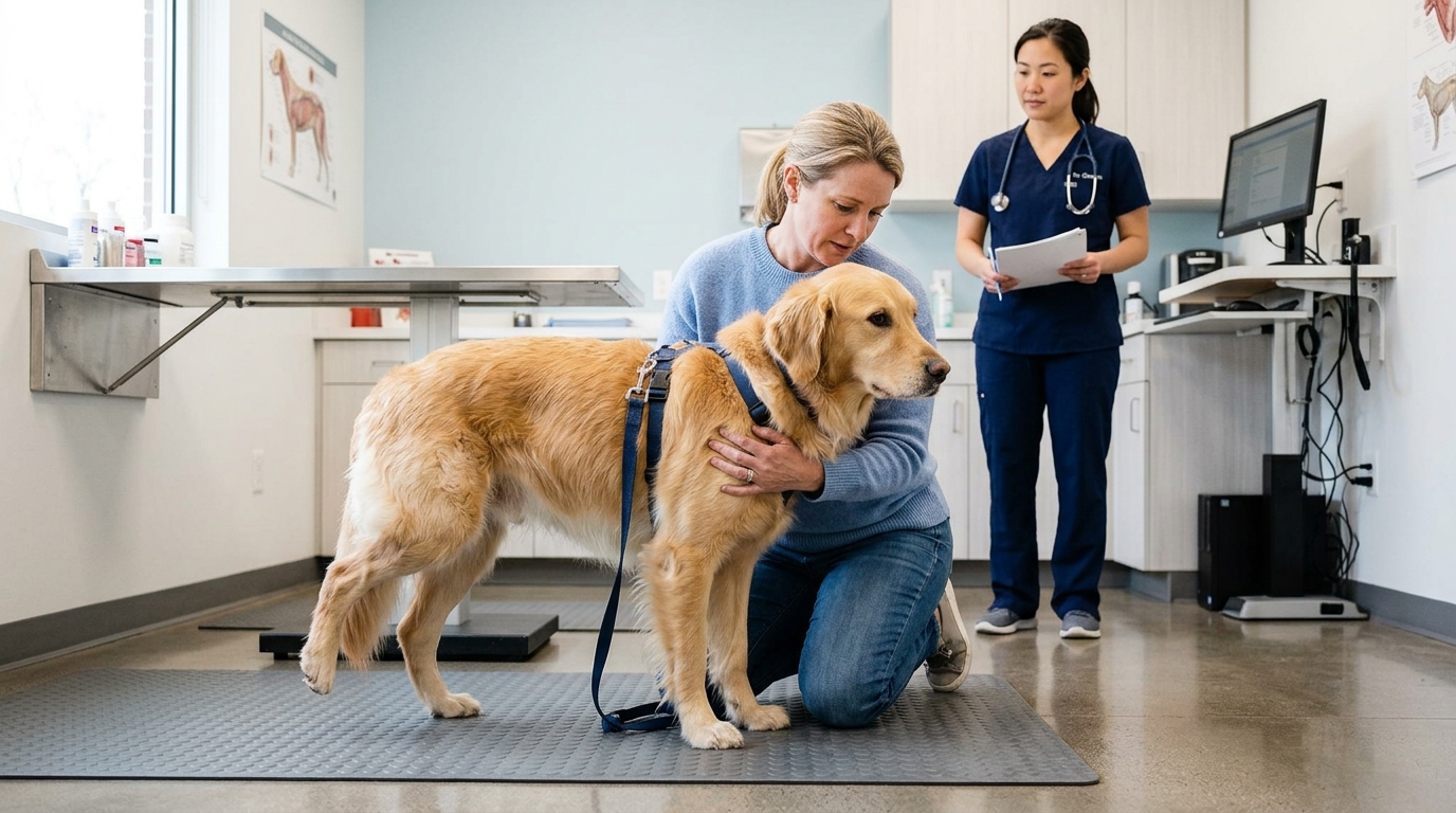
A dog suddenly limping on a back leg — especially after a run, jump, or play session — is one of the most common presentations seen in orthopedic surgery. In many of these cases, the culprit is a ruptured cranial cruciate ligament (CCL), the canine equivalent of the human ACL. It is the single most common orthopedic injury in dogs, and it is one that nearly always requires surgical correction for a good long-term outcome.
What Is the Cranial Cruciate Ligament (CCL)?
The cruciate ligaments are two crossed ligaments inside the stifle (knee) joint that stabilize the joint by preventing the tibia from sliding forward relative to the femur. In dogs, the cranial cruciate ligament (CCL) — anatomically equivalent to the human anterior cruciate ligament (ACL) — is under constant load during standing and movement.
This constant stress, combined with a genetic predisposition in many breeds to ligament degeneration, means that CCL rupture in dogs is often not a single traumatic event but rather the endpoint of a chronic degenerative process. The ligament gradually weakens over months to years before it finally tears. Studies suggest that approximately 40–60% of dogs that rupture one CCL will rupture the other within two years.
Which Dogs Are Most at Risk?
CCL disease is seen in dogs of all sizes and breeds, but certain factors dramatically increase risk:
- Large and giant breeds: Labrador Retrievers, Rottweilers, Newfoundlands, and Mastiffs are among the highest-risk breeds
- Obesity: excess body weight places chronic strain on the ligament
- Neutered dogs: early spay/neuter (before skeletal maturity) has been associated with significantly higher CCL rupture rates in several breeds
- Dogs with steep tibial plateau angles (a conformational factor that amplifies shear forces across the joint)
- Age: peak incidence is in middle-aged dogs (3–7 years), though older and younger dogs are also affected
What Are the Signs of a CCL Tear in Dogs?
The most common presentation is:
- Sudden onset rear limb lameness, ranging from toe-touching to complete non-weight-bearing
- Sitting with the affected leg stuck out to the side (“lazy sit”)
- Swelling or thickening on the inside of the knee joint
- Pain and reluctance to fully extend or flex the stifle
- Muscle atrophy of the affected leg with chronic injury
- A clicking or popping sensation in the knee during movement
With a partial tear, the dog may appear intermittently lame — seeming to improve with rest, then worsening with exercise. This pattern can be misleading, as the partial tear typically progresses to a complete rupture without surgical intervention.
How Is a CCL Tear Diagnosed?
Diagnosis is based on physical examination findings and radiographs (X-rays). The classic diagnostic test is the cranial drawer test and tibial compression test, which demonstrate abnormal forward movement of the tibia when the CCL is ruptured. Sedation or general anesthesia is often needed for accurate assessment.
Radiographs cannot visualize the ligament directly, but they reveal joint effusion (fluid), arthritic changes, and other characteristic findings. Advanced imaging with MRI or diagnostic arthroscopy may be recommended in uncertain cases or to evaluate concurrent meniscal injury.
What Are the Surgical Options?
There are several surgical techniques for CCL repair in dogs. North Springs’ board-certified surgical team performs:
TPLO (Tibial Plateau Leveling Osteotomy): Currently the gold standard for most dogs, particularly large breeds. The surgeon cuts and rotates the tibial plateau to change the geometry of the joint so that the CCL is no longer needed for stability. TPLO has excellent long-term outcomes, with most dogs returning to near-normal function.
Lateral suture procedure: A technique in which a synthetic material is placed outside the joint to mimic CCL function. Generally considered for smaller dogs. North Springs’ surgical team will assess your dog’s size, conformation, and activity level to recommend the most appropriate approach.
Surgery appointments at North Springs are scheduled on a case-by-case basis. For urgent cases where a dog cannot bear weight or is in significant pain, our emergency team is available 24/7 to assess and initiate care.
What Does Recovery Look Like?
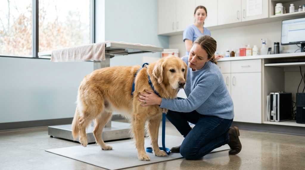
Recovery from TPLO is a structured, multi-week process:
- Weeks 1–2: Strict rest, leash walks only, incision care and pain management
- Weeks 3–8: Gradual increase in controlled leash activity; rehabilitation exercises begin
- Weeks 8–12: Radiographic recheck to assess bone healing; progressive return to activity if healing is confirmed
- 4–6 months: Return to full activity for most dogs
Physical rehabilitation plays a significant role in optimizing recovery. North Springs’ rehabilitation team, led by Dr. Dani Waite, works closely with our surgeons to provide an integrated recovery plan. Services available post-surgery include underwater treadmill therapy, laser therapy, therapeutic exercise, therapeutic ultrasound, electrotherapy, and manual therapy.
When to See a Veterinarian
Any dog with a sudden rear limb lameness lasting more than 24–48 hours, or any dog consistently avoiding weight on a back leg, should be evaluated promptly. Early diagnosis and treatment lead to better long-term outcomes and slower progression of arthritis.
North Springs Veterinary Emergency & Specialty Center’s board-certified surgical team provides comprehensive orthopedic evaluation and surgical treatment. Our rehabilitation team offers post-surgical recovery programs tailored to each patient. Contact us at or call (719) 920-4430 to schedule a surgical consultation. Come to 10520 White Diamond Pt, Colorado Springs, CO or call (719) 920-4430.
Sources and Further Reading
- Witsberger TH, et al. “Prevalence of and risk factors for hip dysplasia and cranial cruciate ligament deficiency in dogs.” JAVMA. 2008;232(12):1818–1824.
- Duval JM, et al. “Breed, sex, and body weight as risk factors for surgical treatment of cranial cruciate ligament rupture in dogs.” JAVMA. 1999;215(6):811–814.
- Wilke VL, et al. “Estimate of the annual economic impact of treatment of cranial cruciate ligament injury in dogs in the United States.” JAVMA. 2005;227(10):1604–1607.
- Griffon DJ. “A review of the pathogenesis of canine cranial cruciate ligament disease as a basis for future preventive strategies.” Veterinary Surgery. 2010;39(4):399–409.
- American College of Veterinary Surgeons. “Cranial Cruciate Ligament Disease.” ACVS.org. https://www.acvs.org/small-animal/cranial-cruciate-ligament-disease
This article is written for educational purposes and does not constitute veterinary medical advice. If your pet is experiencing a medical emergency, seek immediate veterinary care.